PLANET® Onco aims to help Physicians in the implementation of Oncology techniques such as, Diagnosis, Radiation Therapy, Chemotherapy, Adaptive Therapy.
It provides the aid needed with its complete structure segmentations: GTV, BTV, CTV, PTV, OAR (Gross Tumor Volume, Biological Tumor Volume, Clinical Tumor Volume, Planning Target Volume, Organs At Risks) and quantified therapy monitoring from multimodal imaging (CT, MRI, PET, SPECT). The fast, safe, repeatable and non operator dependent features are given to improve data processing.
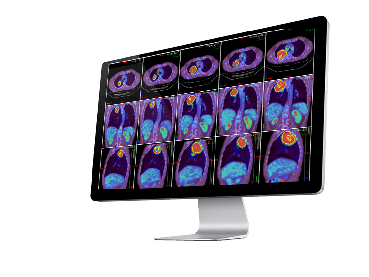
Comprehensive Image Reviewing
- Multi-modality: CT, CTA, CBCT, 3D XA, MRI, PET, SPECT
- Dedicated multi-layout
- Powerful fusion of anatomical and functional series
- Rigid and deformable registration
Advanced Contouring Tools
- Automatic AI-powered segmentation of organs at risk (Auto Segment)1
- Large variety of delineation tools on anatomical and functional modalities
- Planar & multiplanar interpolation capabilities between time points and different modalities
- Interactive modification of contours
- Intensity-based contour delineation (e.g. bones, external body contour extraction)
- 3D margin calculation
Segmentation and Quantification
- One click and automatic detection of lesions (Auto Detect)
- Advanced automatic segmentation on functional image series: Maximum intensity, Nestle, Black, Fitting, Adaptive methods
- Lesion quantification of metabolic activity: volume, SUV mean, SUV max, SUV peak and TLG
- Partial Volume Effect correction
- Specific tool for Sarcoidosis assessment
- Structure agreement quantification (Dice, Jaccard, overlap fraction, sensibility, specificity indices)
Therapy Response Assessment
- Automatic correlation of tumors, segmentation and follow-up across time points
- Disease change assessment using a parametric imaging approach
- RECIST 1.1 and PERCIST 1.0 format reports
Report and Connectivity
- Import & export of image and RT-Struct from/to any DICOM System, TPS
- DICOM Query Retrieve Import/Export (SCP/SCU)
- Integrated report
Radiomics Analysis²
- Computation of first- and second-order features (142 IBSI-compliant indices)
- Intuitive graphical analysis tools (histograms, radar/Kiviat charts, csv sheets…)
- Export capabilities for diagnostics, follow-up & AI-powered radiomics studies
Planet® Onco Dose Edition 3 is developed by DOSIsoft SA in France.
It is a Class IIb CE-marked medical device under MDR (EU) 2017/745 and US FDA 510(k) cleared (not for MAA-planning).
Availability of the product or specific features may vary by country & regulatory approval status. Please contact your local sales representative for more information.
1 This feature uses Total Segmentator 2 Features for research use only